

3D Mammogram
Our state-of-the-art 3D mammography equipment delivers detailed cross-sectional images, enabling your healthcare team to make informed decisions confidently. Whether you’re here for a routine screening or a more specialized diagnostic procedure, our dedicated team is committed to delivering exceptional care and accurate results.
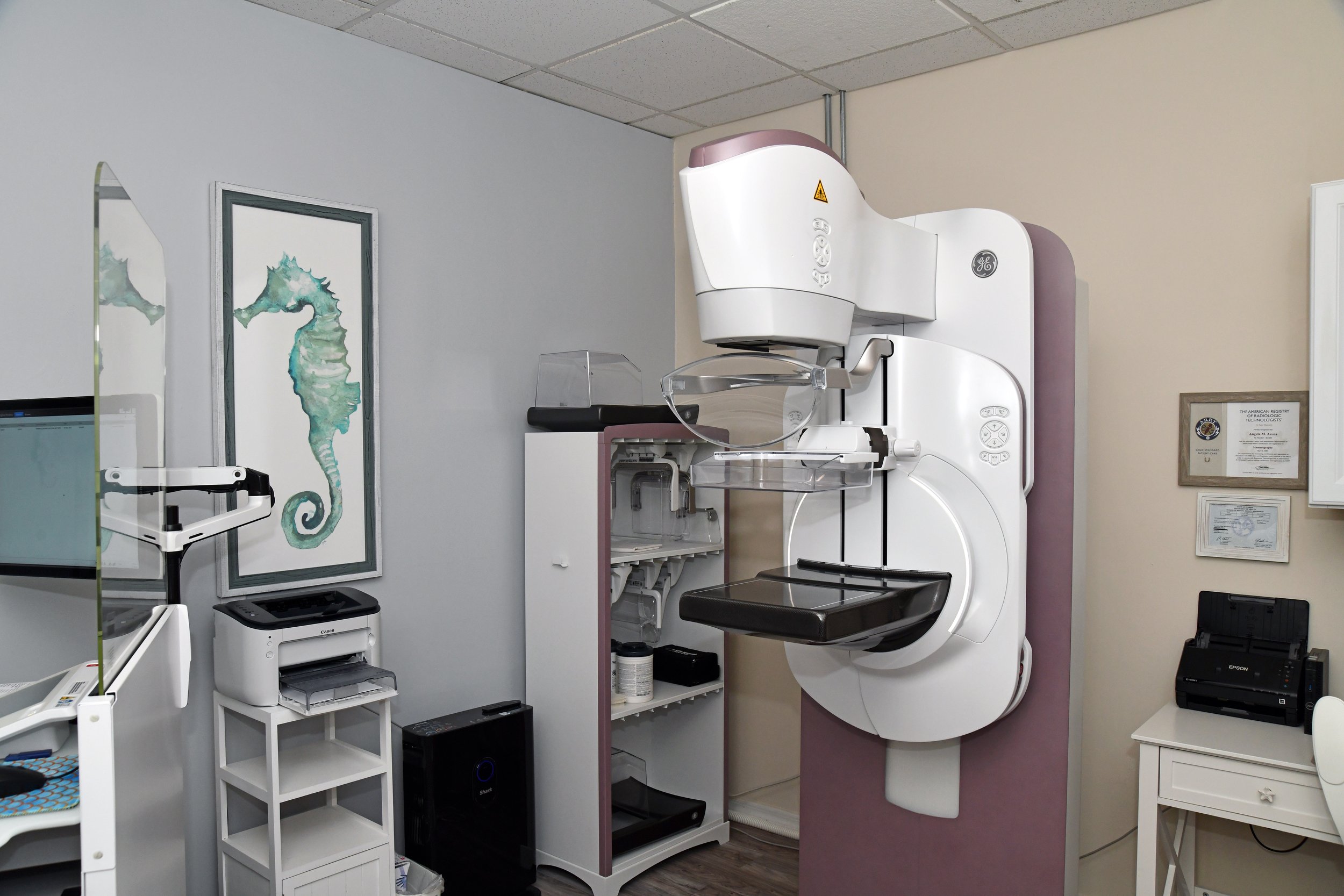
What is a 3D Mammogram?
A 3D mammogram (breast tomosynthesis) is an imaging test that combines multiple breast X-rays to create a three-dimensional picture of the breast.
A 3D mammogram is used to look for breast cancer in people who have no signs or symptoms or to investigate the cause of breast problems, such as breast mass, pain, or nipple discharge.
When used for breast cancer screening, 3D mammogram machines create 3D images and standard 2D mammogram images. Studies show that combining 3D and standard mammograms reduces the need for additional imaging and slightly increases the number of cancers detected during screening.
The 3D mammogram is becoming more common, but it isn’t available at all medical facilities.
Preparing for a 3D Mammogram
Before scheduling a mammogram, the ACS (American Cancer Society) and other specialty organizations recommend that you discuss any new findings or problems in your breasts with your doctor. In addition, inform your doctor of any prior surgeries, hormone use, and family or personal history of breast cancer.
Do not schedule your mammogram for the week before your menstrual period if your breasts are tender during this time. The best time to schedule a mammogram is one week following your period. Always inform your doctor or X-ray technologist if there is any possibility that you are pregnant.
The ACS also recommends:
Before the examination, you will be asked to remove all jewelry and clothing above the waist and given a loose-fitting gown that opens in the front.
Do not wear deodorant, talcum powder, or lotion under your arms or breasts on the exam day. These can appear on the X-ray film as calcium spots.
Describe any breast symptoms or problems to the technologist performing the exam.
Please obtain prior mammogram records when possible and make them available to the radiologist during the current exam.
Ask when your results will be available; only assume the results are normal if you hear from your doctor or the mammography facility.